Publication

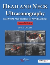
Head and Neck Ultrasonography
Essential and Extended Applications
- Second Edition
- Edited by: Lisa A. Orloff
- Details:
- 544 pages, Color Illustrations (4 Color), Hardcover, 8.5 x 11" 1 lbs
- Included Media:
- Companion Website
- ISBN13:
- 978-1-59756-858-6
- Release Date:
- 04/30/2017
Overview
Head and Neck Ultrasonography: Essential and Extended Applications, Second Edition is a comprehensive text of point-of-care ultrasonography for clinicians who manage patients with head and neck disorders. The Second Edition has been revised to bring the reader up to date in expanded applications of real-time ultrasonography for the spectrum of conditions that affect the head and neck region in adults and children alike.
New to the Second Edition:
- Abundant high-resolution grey scale (B-mode) and color Doppler images throughout
- Augmented chapters on thyroid, parathyroid, salivary gland, and interventional ultrasonography
- New chapters that focus on ultrasound in airway management, pediatrics, global health, and endobronchial procedures
- Special additional chapters on ultrasound documentation, FNA technique, and accreditation
- Liberal use of tables that highlight text material
- Extensively revised throughout to contain current information, guideline recommendations, reviews, and definitions
- A PluralPlus companion website with ample video examples of actual patient examinations
This Second Edition provides new insights, pearls, and practical lessons in ultrasonography for the student of head and neck anatomy, the novice ultrasonographer, and the experienced surgeon or specialist who cares for patients with benign, malignant, or functional disorders of the head and neck.
"Right Trans Thyroid Sonopalpation" video sample from companion website:
Reviews
Liam Flood, FRCS FRCSI, Middlesbrough UK, Journal of Laryngology & Otology (2017):
"This is a substantial textbook, which first appeared in 2008. The second edition adds coverage of such topics as airway management, paediatrics, endobronchial procedures and fine needle aspiration (of which much more anon). The impression is of a surprisingly colourful and well illustrated book, which is remarkable considering the monochrome topic. US does rely on movement, interaction and feedback to the operator, to build up a mental 3D image. Fortunately the countless illustrations are backed with on-line video images. Mind you, an early chapter on emerging advances promises 3D (even 4D) reconstructions, as we have seen in foetal US. There is a multi-author series of chapters covering the basic sciences and normal anatomy, whilst the thyroid obviously dominates the text. For salivary glands and neck masses I increasingly rely on a technology that seemed at a dead end and confined to prenatal checks, just a few decades ago. There are some surprises, with chapters on US of the larynx, the bronchi, the oesophagus, the paranasal sinuses and, especially, the ear where I need some further convincing. Is that Endotracheal tube in the right place? I had not thought of that. Clever. But then comes a series of chapters on interventional US. This is the fun bit. For me, Dr Abele's chapter on "The Science and Art of Optimal Fine Needle Biopsy and Smear Making" made for a marvellous 30 pages, which I have now read three times. Every time I do so, I realise where and in how many ways I have been doing this...if not wrongly, then without that "Art" he describes, at the very least. Use an IV extension tubing, touch the needle on the slide, use a smaller needle (however counterintuitive), try the "snap technique". If I never pick up an US probe in my latter years, this advice has altered my practice. Vascular US, Power Doppler, IV contrast, Shear Wave Elastography will remain the province of the radiologists, but there is so much in this book to appeal to any head and neck oncologist, to the endocrine surgeon, or our vascular fraternity. But it was Chapter 16 that "did it for me" and I almost look forward to my next "Two Week Wait" clinic, even I do lack the US machine, let alone the skill to exploit it, to then guide my needle. There is a real need for a book such as this, to inspire the next generation to develop the skills to perform clinic US, if only at a basic level."John David Cramer, M.D., Northwestern University Feinberg School of Medicine, Doody's (June 2017):
"**Description** This practical book for practitioners is a comprehensive overview of applications of ultrasonography for diseases of the head and neck. This second edition provides a worthwhile update to the 2008 edition, including numerous more complicated applications of ultrasonography in this region. **Purpose** The purpose is to both concisely and systematically summarize applications of ultrasonography for the diagnosis and management of head and neck disorders. Ultrasonography offers tremendous potential, but its application requires practitioners to be knowledgeable about its uses and nuances. This book successfully distills the complexities of ultrasonography in the head and neck into an approachable and practical book for practitioners. **Audience** Intended for practitioners who care for patients with disease of the head and neck, the book is relevant for a broad range of specialists, including otolaryngologists, endocrine surgeons, head and neck oncologic surgeons, endocrinologists, radiologists, pathologists, and trainees in these fields. The book has much to offer practitioners who already use ultrasonography but are interested in mastering the craft and novices. Each chapter is authored by different distinguished specialists from a variety of fields that offer expert perspectives on applications of ultrasonography in the head and neck. **Features** A focus of the book is on thyroid pathology, and for many physicians this will be the most commonly used application, but sections also include basic information on physics and equipment, ultrasonography for salivary glands, neck masses, the larynx, sinuses, vascular assessment, and transesophageal endosonography. New sections include endobronchial ultrasound, airway examination, pediatric applications, and usage in global health. The chapter on the science of optimal fine needle biopsy I found particularly helpful to improve my practice and maximize the diagnostic yield of fine needle aspiration. The book is visually impressive and includes images that vividly depict findings in grayscale as well as color Doppler. **Assessment** This is an exceptional book on head and neck ultrasonography. It succeeds as a practical resource that will enable practitioners to expand their usage of ultrasonography to new areas and maximize their current use."Yasmina C. Ahmed, MD, Bronx, New York, USA, Annals of Otology, Rhinology, and Laryngology, Vol. 126(10) 727 (October 2017):
"Head and Neck Ultrasonography: Essential and Extended Applications (2nd ed) is a comprehensive textbook on office-based and intraoperative head and neck ultrasonography. The second edition has been revised to include up-to-date guideline recommendations and new chapters on ultrasound in airway management, pediatrics, global health, endobronchial procedures, and fine-needle aspiration technique. Dr Lisa Orloff has collaborated with 35 international experts from over 20 institutions across the fields of head and neck surgery, radiology, endocrinology, anesthesiology, neurology, pulmonology, and gastroenterology to create a clinically relevant resource for clinicians who care for patients with head and neck disorders. The book is divided into 21 well-organized chapters written by distinguished specialists with expertise in the given subject and is supported by current and extensive references, including many from otolaryngology journals. Each chapter contains numerous static gray-scale (B-mode) and color Doppler images augmented with supplemental online video clips of actual patient ultrasonography to enhance physician interpretation and performance of ultrasound examinations and procedures. This textbook is intended for clinicians caring for patients with head and neck disease, including otolaryngologists, endocrine surgeons, endocrinologists, radiologists, and pathologists. It will enable practitioners to not only perfect their current use of ultrasonography but also expand their use of ultrasonography to new applications. The chapter on fine-needle biopsy deserves special mention as it successfully distills the subtle nuances of optimal fine-needle aspiration to maximize diagnostic yield."
Preface
Acknowledgments
Contributors
Chapter 1. The History of Head and Neck Ultrasonography
William B. Armstrong and Yarah M. Haidar
Chapter 2. Essential Physics of Ultrasound
Marc D. Coltrera
Chapter 3. Ultrasound Equipment, Techniques, and Advances
Kevin T. Brumund
Chapter 4. Normal Head and Neck Ultrasound Anatomy
Russell B. Smith and Lisa A. Orloff
Chapter 5. Thyroid Ultrasonography
Jennifer A. Sipos and Lisa A. Orloff
Chapter 6. Parathyroid Ultrasonography
Maisie Shindo
Chapter 7. Salivary Gland Ultrasonography
Peter Jecker, Urban W. Geisthoff, Jens E. Meyer, and Lisa A. Orloff
Chapter 8. Ultrasonography of Neck Masses
Hans J. Welkoborsky
Chapter 9. Laryngeal Ultrasonography
Timothy J. Beale, Lisa A. Orloff, and John S. Rubin
Chapter 10. Transesophageal Endosonography
Michael Mello and Thomas J. Savides
Chapter 11. Endobronchial Ultrasound
Mihir S. Parikh and Ganesh Krishna
Chapter 12. Airway Ultrasonography and Its Clinical Application to Airway Management
Lena Scotto, Lynn Cintron, Fredrick G. Mihm, and Vladimir Nekhendzy
Chapter 13. Head and Neck Ultrasound in the Pediatric Population
Veronica J. Rooks and Benjamin B. Cable
Chapter 14. Ultrasonography of the Face, Paranasal Sinuses, and Ear
Peter Jecker
Chapter 15. Interventional Ultrasonography
Vinay T. Fernandes and Lisa A. Orloff
Chapter 16. The Science and Art of Optimal Fine Needle Biopsy and Smear Making
John Stephen Abele
Chapter 17. Office-Based Ultrasonography: Documentation and Reporting
Ilya Likhterov, Meghan E. Rowe, and Mark L. Urken
Chapter 18. Vascular Ultrasound, Carotid, and Transcranial Doppler Imaging
Tatjana Rundek, Marialaura Simonetto, Nelly Campo, and Digna E. Cabral
Chapter 19. Ultrasound Surveillance of the Neck in Head and Neck Cancer
Michiel W.M. van den Brekel and Jonas A. Castelijns
Chapter 20. Head and Neck Ultrasonography in Global Health
Merry E. Sebelik
Chapter 21. Head and Neck Ultrasound: Certification and Accreditation
Lisa A. Orloff
Index
About The Editor
Lisa A. Orloff, MD, FACS, FACE, is Director of the Endocrine Head and Neck Surgery Program and Professor of Otolaryngology - Head and Neck Surgery at Stanford University School of Medicine and the Stanford Cancer Center. Her clinical practice has evolved from a long and broad emphasis in head and neck oncology, laryngology, and microvascular reconstructive surgery to a more specific focus on the surgical management of thyroid and parathyroid tumors. Dr. Orloff served three consecutive terms as the Chair of the American Academy of Otolaryngology-Head and Neck Surgery (AAO-HNS) Endocrine Surgery committee. She holds leadership roles within the American Head and Neck Society, the American Thyroid Association, the American Institute of Ultrasound in Medicine, the American Association of Clinical Endocrinology, and the American College of Surgeons. She is co-chair of the Thyroid, Parathyroid, and Neck Ultrasound training program at the ACS and a member of the ACS National Ultrasound Faculty. Dr. Orloff is a former Fulbright scholar, and she has been a voting member of the U.S. Food and Drug Administration's panel to evaluate medical devices for otolaryngology.
Related Titles
Stroboscopy
408 pages, Color Illustrations (4 Color), Hardcover, 8.5 x 11"
Atlas of Laryngoscopy
Robert Thayer Sataloff, Mary J. Hawkshaw, Johnathan B. Sataloff, Rima A. DeFatta, Robert Eller
368 pages, Color Illustrations (4 Color), Hardcover, 8.5 x 11"
Parathyroid Surgery
Edited by: David Terris, William S. Duke, Janice L. Pasieka
248 pages, Color Illustrations (4 Color), Hardcover, 8.5 x 11"
Head and Neck Vascular Anomalies
Gresham T. Richter, James Y. Suen
456 pages, Color Illustrations (4 Color), Hardcover, 8.5 x 11"
Contemporary Transoral Surgery for Primary Head and Neck Cancer
Edited by: Michael L. Hinni, David G. Lott
264 pages, Color Illustrations (4 Color), Hardcover, 8.5 x 11"
Atlas of Otoscopy
Joseph B. Touma, M.D., F.A.C.S., B. Joseph Touma, M.D.
208 pages, Color Illustrations (4 Color), Hardcover, 8.5 x 11"
Purchasers of this book receive complimentary access to supplementary materials hosted on a PluralPlus companion website.
Materials:
- Companion videos
To access the materials, log in to the website using the URL and Access Code located inside the front cover of your copy of Head and Neck Ultrasonography, Second Edition.


